Friday, November 18, 2011

Day 36. Thurs Nov. 17, 2011.

This week it took me forever to find any life in my tank. I searched under the microscope for a while before stumbling upon anything lively. It was more Petelomonas. I snapped another picture just for the sake of the blog (Figure 1).

Figure 1. Petalomonas (Patterson 1996, Fig. 83).
I also found more Tachyosoma. They were lively, just like the first time I saw them. I'm curious why they disappeared between lab visits. Maybe we misidentified Euplotes last time? Figure 2 is an image of Tachyosoma and Phacus triqueter, and Figure 3 is an image of Phacus triqueter alone.
Figure 2. Tachysoma (Patterson 1996, Fig. 264-265).
Figure 3. Phacus triqueter (Forest 1954, Fig. 419).

Sources:
Forest, Herman Silva. 1954. Handbook of Algae. Knoxville (TN): University of Tennessee Press. 296 p. Fig. 419. 

Patterson, D. J. 1996. Free-Living Freshwater Protozoa: A colour guide. London (England): ASM Press. 55-125 p. Fig. 83, 264, 265.

Thursday, November 10, 2011

Day 27. Tues Nov 8, 2011

This week there was much less diversity and activity in my aquarium. It actually took me a few minutes to even find any organisms that moved. However, even though there was less diversity within my aquarium, there were plenty of organisms for me to identify. The Tachyosoma from the week before were gone, but there was another organism that greatly resembled them. In fact, I mistook it for the Tachyosoma, but Dr. McFarland assured me it was not the same organism. I identified it as Euplotes (Patterson 1996). These creatures crawl instead of swim, according to Free-Living Freshwater Protozoa: A colour guide. The organism is shown in Figure 1.
Figure 1. Euplotes (Patterson 1996, Fig. 260).

This was not, however, the first organism that I noticed in my aquarium. I saw Bursaria first but was not able to get a picutre (it quickly swam away and I didn't find it again). It was a some-what large organism with a long flagella. Among the other organisms I identified was Paramecium: A fat grub-like organism that moved really slowly (Patterson 1996). It is viewable in Figure 2.
Figure 2. Paramecium (Patterson 1996, Fig. 346).
Litonotus, pictured in Figure 3, was a smaller looking version of Paramecium and swam a little faster (Patterson 1996).
Figure 3. Litonotus (Patterson 1996, Fig. 285).
Next I identified Petalomonas (Patterson 1996). This organism was more similar to the Bursaria, but it was much smaller and not nearly as fast. There were a lot of these guys, but capturing an image that illustrated them well was a challenge. Figure 4 was my best attempt.
Figure 4. Petalomonas (Patterson 1996, Fig. 83).
One of the neatest organisms that I found was a free-swimming algae called Phacus triqueter (Forest 1954). It stood out from the rest of the algaes and even the protozoa. It was a shade of green that differed from every thing around it. The way it swam was cool too. It was spinning as it swam, so it looked almost like a coin being flipped (It was a flat organism, hence the resemblance of a coin). Figure 5 doesn't show the organism's shape well, but it was the best image that I could manage.
Figure 5. Phacus triqueter (Forest 1954, Fig. 419).

Sournces:
Forest, Herman Silva. 1954. Handbook of Algae. Knoxville (TN): University of Tennessee Press. 296 p. Fig. 419.

Patterson, D.J. 1996. Free-living Freshwater Protozoa: A Colour Guide. London (England): ASM Press. 55-154 p. Fig. 83-346.

Wednesday, November 2, 2011

Day 20. Tues. Nov 1, 2011.

There were a lot more organisms on my most recent visit to the lab, so many that I couldn't take the time to name them all! The very first thing I noticed when I placed my aquarium under the microscope (before I even turned on the light) was a little organism swimming very fast through the water. I could SEE this thing with my eye, so I knew it was something cool. It was really hard to get a picture because as soon as I'd get the lens focused it would swim away! However, I did manage to snap a decent shot. Figure 1 is an image of Tachysoma (Patterson 1996). I don't remember exactly what family this organism falls into, but I think  Dr. McFarland mentioned that it might be a Cyclops. As it turns out, the one I took a picture of was undergoing cell division. I also identified an organism called Peranema, viewable in Figure 2 (Patterson 1996). It is a Euglenid. These types of organisms have flagella that flap and move them forward. There were more of these, but most of them were very very tiny (They were small on the 40 objective). The only other organism that I identified on my last visit was called  Nostoc (Lund 1995). It's body has little notches between cells, and it slowly swims through the water. These organisms usually live in colonies made of filaments that form a gelatinous sheath around them, but this one, shown in Figure 3, was alone. My guess is that I will see some of these algae colonies in the near future.

Figure 1. Tachyosoma (Patterson 1996, Fig. 264-265).


Figure 2. Paranema (Patterson 1996, Fig. 70-72).


Figure 3. Nostoc (Lund 1995, Fig. 445-447).
Sources:
Lund, J.W.G. 1995. Freshwater Algae: Their Microscopic World Explored. England: Biopress Limited. 234 p. Fig.445-447

Patterson, D.J. 1996. Free-living Freshwater Protozoa: A Colour Guide. London (England): ASM Press. 51 p. Fig. 70-72.

Tuesday, October 25, 2011

Day 13. Tues Oct 25, 2011

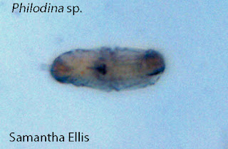
Figure 1. Philodina (Smith 2001, Fig. 7.11).
It has been 13 days sense I last saw my aquarium. One food pellet was added on the 21st (The food pellet information is listed below). At first, I didn't see much of anything in my aquarium. Not even anything near the food pellet. Once I got closer to the sediment I began to see a few organisms. Most of them were too small for me to identify, but one organism was very obvious. I identified it as Philodina, which is a Rotifer. Rotifers are omnivores, meaning they eat virtually anything (Smith 2001). This organism was very interesting. For a while it stayed in the same spot and kept moving its jaws back and forth like it was chomping on something, but then suddenly swam away. I tried to follow it with the microscope but I couldn't keep up, and I never found it again.

Figure 2. Philodina's movement (Smith 2001, Fig. 7.11).
Sources:
Smith, D.G. 2001. Pennak's Freshwater Invertebrates: Porifera to Crustacea. 4th ed. Canada: John Wiley and Sons. 139 p. Fig. 7.11.

Beta Food Ingredients: "Atison's Betta Food" made by Ocean Nutrition, Aqua Pet Americas, 3528 West 500 South, Salt Lake City, UT 84104. Ingredients: Fish meal, wheat flower, soy meal, krill meal, minerals, vitamins and preservatives. Analysis: Crude Protein 36%; Crude fat 4.5%; Crude Fiber 3.5%; Moisture 8% and Ash 15%

Wednesday, October 12, 2011

Day 1. Wed Oct 12, 2011

Today we made our micro aquariums! I chose the water from the plastic bird bath mainly because it was behind me and I didn't feel like traveling to obtain water from another source. Lazy, I know. We added two species of plant so that organisms could have something to gather around or eat. They were Amblestegium (Plant A) and Utricularia gibba (Plant B). I looked at my aquarium under the microscope for a little while, but my water didn't contain many organisms, so I stopped a little earlier than others did. I did see what looked like a nematode and a few black and white specs that seemed to be swimming, and Ken showed me some sort of bladder on one of the sample plants that we added. I'm really excited to see what kinds of changes will occur over the course of this experiment. I hope more organisms will inhabit my aquarium because it's just so neat to watch them swim around!

Water Source: Plastic Bird Bath pool . 0.9 mile from Fountain City Pond on Fountain Rd. Knox Co. Knoxville TN Partial shade exposure N 36o02.249' W083o55.999' 1121 ft 10/9/2011

Plant samples: Plant A . Amblestegium sp. Moss. Collection from: Natural spring. at Carters Mill Park, Carter Mill Road, Knox Co. TN. Partial shade exposure. N36 01.168 W83 42.832. 10/9/2011

                      Plant B. Utricularia gibba L. Flowering plant. A carnivous plant. Original material from south shore of Spain Lake (N 35o55 12.35" W088o20' 47.00), Camp Bella Air Rd. East of Sparta Tn. in White Co. and grown in water tanks outside of greenhouse at Hesler Biology Building. The University of Tennessee. Knox Co. Knoxville TN